The France Life Imaging infrastructure is organized into 4 thematic expertise networks – molecular imaging agents, instrumentation and technological innovation, interventional imaging and multimodal imaging processing and analysis to harmonize research in key biomedical imaging fields – and a training network.
Networks of expertise
1
RE1 – Molecular imaging agents
Presentation
Associated laboratories:
Cobra Laboratory – Organic Chemistry Team, Rouen , ICMUB P2DA team, Dijon
The production of imaging agents has been identified as a major obstacle to medical imaging research. Although some laboratories have the capacity to generate these agents for their own investigative work, they are not in a position to supply other centers. As a result, the aim of this workpackage is to provide a framework for the production of imaging agents for which there is no commercial supply, and to generate new generate new imaging probes or contrast agents for in vivo for in vivo biomedical research and clinical applications (e.g. optical agents, radionuclides, magnetic resonance agents, etc.).
The RE1 aims to stimulate interdisciplinarity in collaborative medical imaging projects, and accelerate the design, manufacture and eventual clinical use of new molecular imaging agents with greater sensitivity, selectivity and minimal toxicity.
RE1 is thus organized into 4 sub-modules:
WP1.1: Modular targeting platform for imaging
Development of synthetic macromolecules & marking techniques
WP1.2: development of probes and contrast agents
Introduction of short-lived radionuclides
Optimizing the binding properties of chelating agents (for applications in MRI, Optics, SPECT and PET)
Optimization of agents used in MRI and optical imaging
Development of innovative technologies including clickable, intelligent and multimodal probes
WP1.3: nanoparticles for imaging
– development of nanoparticles for multimodal imaging
– development of multifunctional nanoparticles with high specificity and detectability in imaging
WP1.4: Proof of concept, clinical applications, translational research
Scale up process: from the chemistry lab to production
- Proof of concept and preclinical and clinical trials
2
RE2 – Technological innovations and instrumentation
Presentation
Medical imaging has been an established part of the clinical arsenal for over 30 years. Instrumental and technological developments continue to be the main driving force behind research aimed at better establishing the presence or severity of disease, and assessing response to treatment using more relevant clinical and preclinical information.
The aim of RE2 is to develop and promote specific approaches and advances to enable breakthroughs in each imaging modality. Beyond these individual challenges, developments combining two or more modalities open up radically new perspectives in diagnosis and therapy, with enhanced sensitivity and specificity.
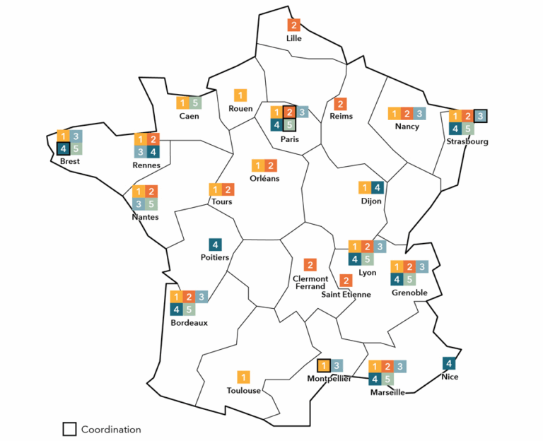
Associated laboratories
3
RE3 – Interventional Imaging
Presentation
RE3 research centers :
| Laboratoires | City | Hub |
| BioMaps | Orsay | Hub Paris Sud |
| C2N | Saclay | Hub Paris Sud |
| CERIMED | Marseille | Hub Marseille |
| CERMEP | Lyon | Hub Lyon |
| Bordeaux University Hospital | Bordeaux | Hub Bordeaux |
| Poitiers University Hospital | Poitiers | |
| Clinatec | Grenoble | Hub Grenoble |
| CRAN | Nancy | Hub Grand Est |
| CRCL | Lyon | Hub Lyon |
| CREATIS | Lyon | Hub Lyon |
| CRI | Paris | Hub Paris Centre |
| CRMBM | Marseille | Hub Marseille |
| CRNL | Lyon | Hub Lyon |
| FEMTO-ST | Besançon | |
| GIN | Grenoble | Hub Grenoble |
| GREMAN | Towers | |
| Tenon Hospital | Paris | Hub Paris Centre |
| IADI | Nancy | Hub Grand Est |
| Icube | Strasbourg | Hub Grand Est |
| IHU Lyric | Bordeaux | Hub Bordeaux |
| IHU Strasbourg | Strasbourg | Hub Grand Est |
| Imaging and Brain | Towers | |
| IMNC | Orsay | Hub Paris Sud |
| INCIA | Bordeaux | Hub Bordeaux |
| Langevin Institute | Paris | Hub Paris Centre |
| Pasteur Institute | Paris | Hub Paris Centre |
| IPHC | Strasbourg | Hub Grand Est |
| IRCAD | Strasbourg | Hub Grand Est |
| IRISA | Rennes | Hub Grand Ouest |
| IRIT | Toulouse | Hub Occitan |
| ISCR | Rennes | Hub Grand Ouest |
| Biochemistry laboratory | Angers | |
| LabTAU | Lyon | Hub Lyon |
| LaTIM | Brest | Hub Grand Ouest |
| LCMCP | Paris | Hub Paris Centre |
| LEM3 | Nancy | Hub Grand Est |
| LIB | Paris | Hub Paris Centre |
| LIIE | Marseille | Hub Marseille |
| LIRMM | Montpellier | Hub Occitan |
| LISE | Paris | Hub Paris Centre |
| LISSI | Vitry sur seine | |
| LMA | Marseille | Hub Marseille |
| LTSI | Rennes | Hub Grand Ouest |
| MIG-CHU Nimes | Nimes | |
| Neurospin | Gif sur Yvette | Hub Paris Sud |
| Physics for Medicine | Paris | Hub Paris Centre |
| RMSB | Bordeaux | Hub Bordeaux |
| TIMC | Grenoble | Hub Grenoble |
Medical and interventional imaging is a major challenge for better understanding, diagnosing, predicting and curing pathologies (neurological, oncological, cardiological, vascular, etc.).
Imaging-controlled interventional procedures are expanding rapidly, and could overtake surgical procedures within the next 10 years. This increase is closely linked to technological advances in fields ranging from imaging systems and image processing to interventional devices and robotic systems.
The aim of interventional radiology is to treat a variety of pathologies (tumors, vascular lesions, etc.) using so-called “minimally invasive” techniques guided by intraoperative imaging, with access via natural channels, blood vessels or the percutaneous route.
Interventional imaging is characterized by the development of intra-operative imaging techniques (in the broadest sense) to assist medical-surgical procedures, in order to improve the quality of care and shorten hospital stays.
This rapidly evolving field lies at the frontier between several medical specialties: surgery, radiology, endoscopy, radiotherapy, cardiology/neurology…
The RE3 FLI takes several approaches to this theme:
WP3.1: navigation and augmented reality
- Development of algorithms for superimposing pre-operative data on intra-operative images in a deformable, moving environment
- Development of algorithms for non-rigid fusion of patient images from different imaging modalities
- Development of robust registration algorithms for navigation applications without the need for patient markers and landmarks
- Development of models of the surgical procedure (e.g. based on large volumes of data from procedures already performed)
- Development of algorithms to recognize surgical activities in the operating room
- Development of algorithms for visual tracking and recognition of interactions between tools and anatomy
- Development of decision-support tools in the OR control tower
– new context-sensitive user interfaces
– reminders (call next patient) and alerts (anomalies)
– optimization of OR management - Development of new context-sensitive radiation protection methods that take into account the 3D layout of the room (positioning of clinicians and equipment, C-arm parameters).
- Real-time surgical guidance
- Development of real-time 3D planning and monitoring techniques for conformal therapy using physical agents (e.g. HIFU – High Intensity Focused Ultrasound)
- Monitor and integrate emerging technologies and innovations in the field of augmented reality, both for interventional scene acquisition and multimodality information fusion representation (pre- and intra-operatively).
Develop AR/IA methodologies, in particular to optimize the guidance of interventional procedures.
WP3.2: Robotic imaging and intervention systems
- Robotization of flexible endoscopes for endoluminal and transluminal operations
- Development of collaborative and semi-automated assistance modes (including visual servoing)
- Image-based visual servoing for physiological motion compensation and fine control of instrument movements
- Intraoperative medical imaging-guided robotic procedures
- Control of complex movements inaccessible to the human hand
- Expertise in robotic instrument-tissue interactions (confocal endomicroscopy, catheterization, etc.)
- Robotic retrocontrol of HIFU therapy to optimize energy deposition in target tissues based on monitoring imagery (3D scanning, 3D mechanical motion compensation)
- Needle gripping and insertion
WP3.3: Magnetic resonance imaging-guided interventions
- Modeling MRI-guided surgical procedures
- Optimization of real-time MRI sequences for instrument guidance
- Quantitative MRI for procedure monitoring (elastography, temperature, etc.)
- Development of motion compensation algorithms
- Development of MRI-compatible instruments for interventional radiology
- Fine control of robots for MRI gestures
- Real-time object tracking under MRI
WP3.4: Ultrasound-based therapies (HIFU and targeted vectorization)
- Development of HIFU systems for new clinical indications (excluding uterine fibroids)
- Development of real-time temperature monitoring systems for HIFU therapy control
- Ultrasound beam control (HIFU) for motion planning and/or compensation in therapy
- Development of intracorporeal systems for HIFU therapy of deep-seated targets
- Ultrasound biomodality: US imaging (ultrasound)/HIFU therapy
- Development of LIPUS (Low Intensity Pulsed Ultrasound) and LEUS (Low Energy Ultrasound) for new clinical indications (neurodegenerative diseases/disabilities of neurological origin, cardiac arrhythmias, drug delivery, immunotherapy, neuro/cardio-protection).
- Development of US 4D therapy (3D + real-time)
- Development of 4D planning and monitoring techniques for conformal HIFU therapy
- Development of registration/fusion techniques between medical imaging data and modeled acoustic/thermal data for HIFU therapy
WP3.5: new intraoperative and endoscopic imaging modalities
- Development of imaging modalities compatible with operating constraints for intraoperative or endoscopic monitoring of surgery
- Development of robust techniques for intraoperative or endoscopic detection of pathological conditions (e.g. cancer, margins, lymph nodes, hypoxia) or vital structures (e.g. vessels, nerves, ureters).
- Development of intraoperative or endoscopic biopsy techniques using imaging or spectroscopy
- Innovative percutaneous procedures (US / X-ray guidance)
- Endomicroscopy (Confocal, OCT…)
- Improved medical imaging (functional and/or structural contrast)
- Quantitative, multimodal and multiscale approaches
- Development of real-time tissue characteristic monitoring systems for HIFU therapy monitoring (US thermometry, US elastrography)
- Development of US 4D imaging
- Development of quantitative US imaging (QUS)
WP3.6: Image-guided radiotherapy
- Improved therapeutic targeting
- Improvement and control of dose distribution and deposition
4
RE4 – Multimodal Imaging Processing and Analysis
Presentation
Research centers :
LaTIM, CREATIS, IPHC-ImaBio, LTSI, ICUBE, CERMEP, CEA SHFJ, Visages, GIN, LaBRI, INCIA, CRMBM, CPPM, CRNL, Télécom ParisTech
Multimodal imaging is playing an increasingly important role in diagnosis and therapeutic follow-up. This is made possible in part by
In order to coordinate scientific work in clinical and pre-clinical image processing at a national level, the ER4 has set up 5 sub-groups:
WP4.1 : Reconstruction of mutlidimensional images
Parametric modeling, model selection and regularization techniques
Incorporation of motion models in reconstruction phases
WP4.2: Multimodal image processing
Precise determination, extraction and exploration of image-derived parameters
development of deformable space-time fusion models, organ reconstruction and patient personalization
WP4.3: Databases, data mining, knowledge modeling
Development of methodologies for database construction (interoperability and management of data heterogeneity), individual patient characterization, automatic classification,
development of methods for creating atlases of pathology models and their mechanisms
WP4.4 : Numerical simulations
Construction of clinically realistic simulation databases (pathology models and their mechanisms, intra-patient anatomical and functional variability, etc.).
WP4.5 : Signal processing in MEG / EEG
Modeling the functional connectivity of the brain network,
development of multimodal MEG/EEG,
development of new research and clinical applications of real-time electrophysiology,
development of methods to avoid, correct and eliminate artifacts,
development of methods for MEG-based reconstruction,
implementation and validation of protocols to meet ISO 9001 standards
5
RF – Training
Presentation
Background
Rapid and promising advances in biomedical imaging have highlighted the need to train a new generation of professionals, evolving in a multidisciplinary environment and thus requiring knowledge of biology, chemistry, fundamental physics, image analysis and clinical applications. The training of future imaging professionals is a real challenge, given the diversity of imaging modalities, particularly multimodal imaging, which considerably increases the potential of imaging, and the challenge of data analysis. In this context, the training of users and platform staff is essential.
Coordination
The coordination of training initiatives within the FLI network will not only help meet the need for qualified manpower to meet the new challenges facing the industry, but also ensure the sustainability, professionalism and recognition of these training courses. This mission will be carried out by the CEA’s Institut National des Sciences et Techniques Nucléaires CEA’s Institut National des Sciences et Techniques Nucléaires, which has set up a Steering Committee comprising representatives from the various nodes, as well as representatives from off-node technology platforms and learned societies.
The objectives
Ensure a high-level training offer for students and professionals (academic and industrial) in the field of biomedical researchAssure the visibility of this training offer by stimulating exchanges within the scientific imaging community and strengthening existing networks
Ensuring the sustainability of this training offer
Ensure professional recognition of these training courses (in France and eventually in Europe).
The missions
Optimizing training for imaging students
Identify and publicize existing training courses
Contribute to the implementation of innovative TP on platforms
Organize and support Summer Schools or Workshops linked to the various nodes
Encourage student mobility for training purposes by providing financial support
Contribute to the professional integration of students by creating a national office in charge of student employability.
Coordinating continuing education in imaging
Identify continuing training needs
Design and coordinate new training courses for the community, platform staff and users
Promoting continuing education
Strengthen continuing education offerings for industrial partners
Relay job opportunities/offers on platforms via the website
Stimulating and financing inter-platform staff mobility
Deployon an international scale imaging training programs
Organize English-language training sessions
Promote international exchanges by financing the participation of staff and students in conferences and training courses outside France, and by inviting international experts.