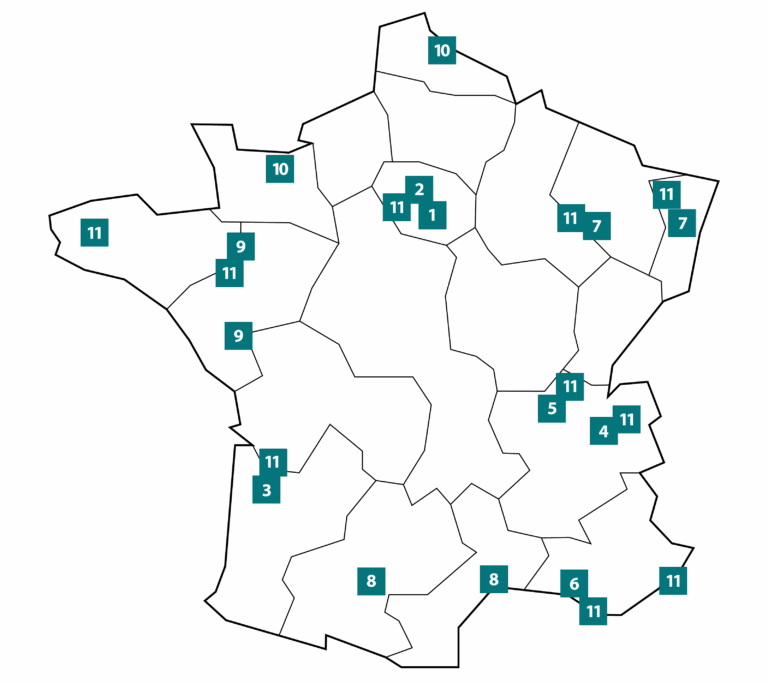
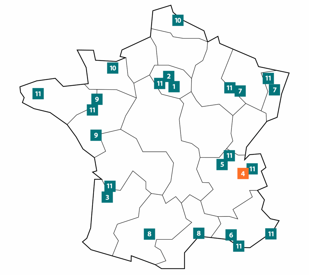
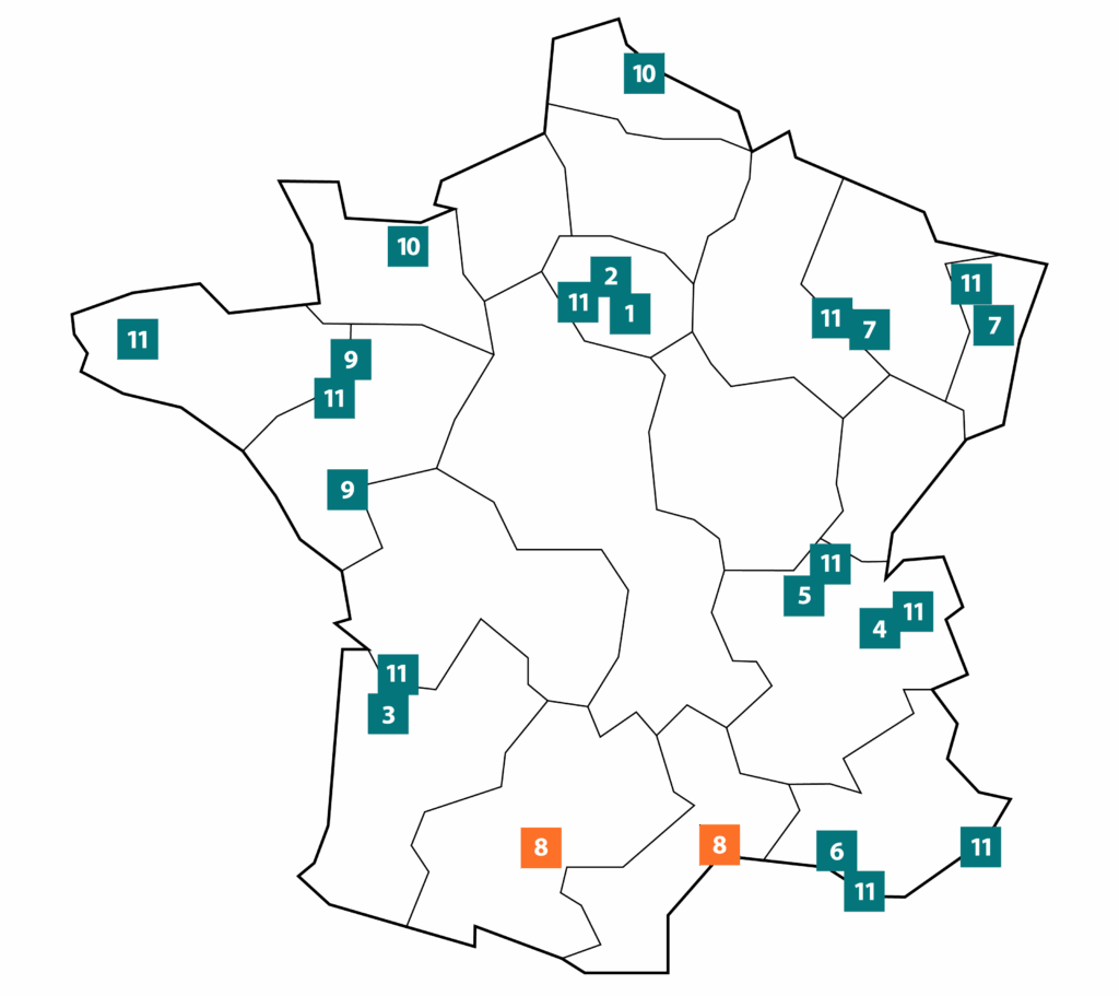
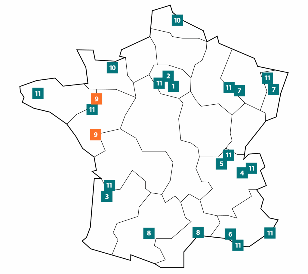
The France Life Imaging infrastructure comprises 10 regional hubs and one transverse hub, all of recognized scientific and technological quality and committed to a partnership approach.
The hubs
- if the platform is geographically close to a hub, it can integrate this hub
- if several platforms in the same region and/or in related regions form a coherent whole that has already been accredited, they can form a new regional hub of imaging platforms.
Depending on the case, FLI offers two procedures, both of which can be downloaded:
- the application procedure for a platform to join an existing hub, Elargissement FLI – PF
- the application procedure for a new hub to join the network, Elargissement FLI – Hub vf
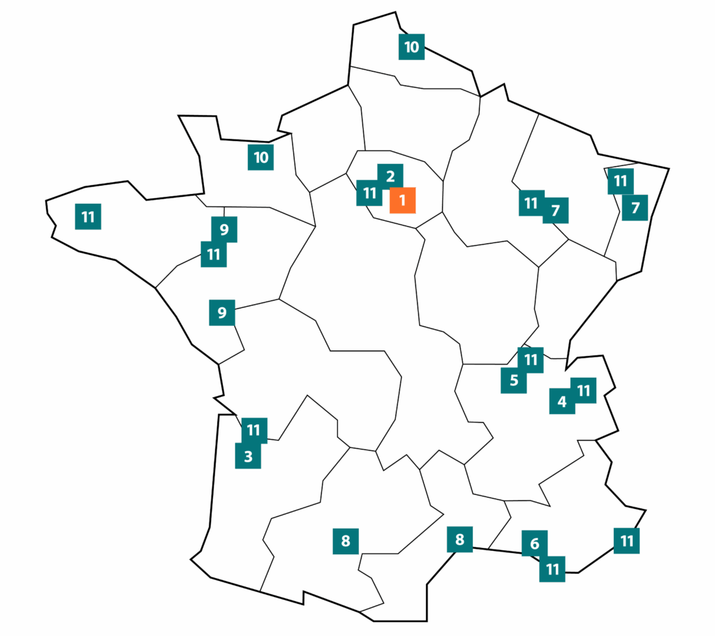
1
Paris Sud
Presentation
Part of the Paris-Saclay Campus, the Paris-Sud hub brings together over 200 researchers and technicians in three biomedical imaging research units (UMR BioMaps hosted by CEA/SHFJ, UMR Baobab hosted by CEA/NeuroSpinet UMR 8165) located in Orsay, Saclay, Villejuif and Kremlin-Bicêtre. The node brings togetherleading methodological expertise in the main medical imaging modalities: MRI, nuclear imaging (PET instrumentation and radiolabeling of molecules), ultrasound and optical imaging. Expertise in digital simulation, image processing and compartmental modeling completes this know-how. The main specificity of the Paris Sud node is the strong involvement of the physics community, thanks to its location at the heart of a unique Physics/Technology ecosystem (Orsay campus / Saclay research center). Transfer to the clinic focuses mainly on brain diseases (neurology, psychiatry) and oncology, in conjunction with university hospitals in the south of Paris.
The node offers an exceptional environment of imaging platforms featuring advanced techniques in nuclear imaging (PET-MRI, high-resolution brain PET), MRI (world’s only 17T preclinical and forthcoming 11.7T clinical systems), ultrasound imaging (ultrafast ultrasound) and optical imaging (biphotonics), complemented by MEG and EEG.
Core imaging expertise and know-how
The node’s expertise covers :
- Ultra-high-field MRI, in particular the development of an in vivo ultra-high-field MRI system for mapping cognitive functions at several scales (microscopic, mesoscopic and macroscopic);
- The development of radiopharmaceuticals for PET;
- Signal and image processing, with digital simulation, tomographic reconstruction, multi-modality image processing and statistical analysis. This area involves most of the imaging laboratories associated with Université Paris Saclay;
- a multidisciplinary environment combining physics, mathematics and engineering, conducive to the development of tomorrow’s medicine, with enhanced sensitivity and specificity, and easier access to patients;
- population-based studies, conducted in particular as part of the CATI program (neuroimaging by MRI and PET), integrated into the constitution of a large cohort of subjects recruited on the basis of cognitive complaint criteria, and also as part of the European “Human Brain Project” program;
- Intraoperative anatomopathological analysis based on two-photon microscopy.
Preclinical and clinical imaging platforms / responsible :
- Molecular and multi-modal imaging: Service Hospitalier Frédéric Joliot SHFJ – Vincent Lebon
- Preclinical ultrasound imaging: Gustave Roussy – Nathalie Lassau
- Preclinical optical imaging: Gustave Roussy – Corinne Laplace-Builhé
- Two-photon imaging : IJCLab, pole physique-Santé- Darine Abi Haidar
Hub laboratories:
- UMR BioMaps, UMR9012
- IJCLab, Health Physics Center
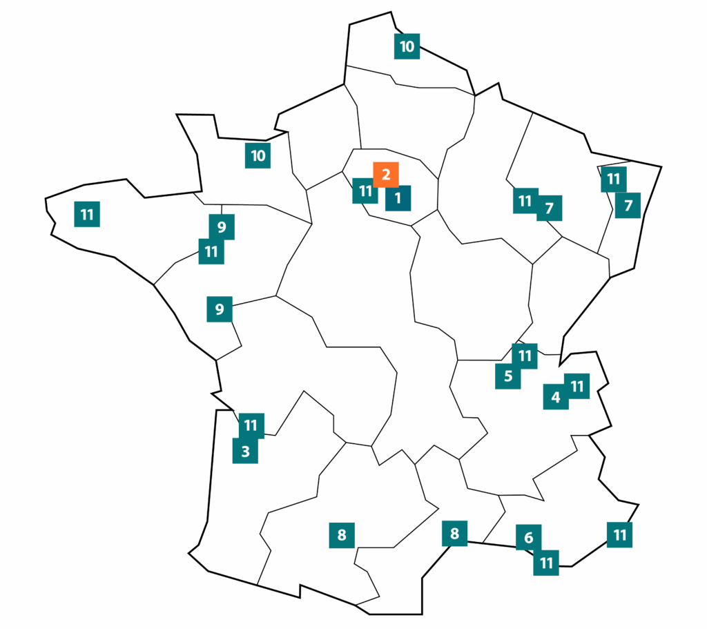
2
Paris Centre
Presentation
The Paris Centre hub is made up of 4 major research centers integrated into the University of Paris and Sorbonne University, all located in the vicinity of university hospitals (HEGP, Sainte-Anne, Cochin, Bichat-Beaujon, Pitié-Salpêtrière). This proximity facilitates the development of transfer research focused on patient care. This is underpinned by the teams’ multimodal imaging expertise (MRI, PET, SPECT, US, Optical, CT, MEG-EEG, EPR, Magnetic Resonance Elastography and Ultrasound) and integrates several levels, from fundamental research in cognitive neuroscience and biomedical imaging methodology to in vivo imaging of small animals, clinical research and diagnosis and therapeutic follow-up of patients.
Key imaging expertise and know-how developed as part of FLI’s scientific activities
Imaging biomarkers are at the heart of the Paris Centre node’s research, the aim being to develop effective tools for diagnosis and for assessing prognosis and response to treatment. The research teams focus primarily on pathologies such as cancer, inflammation, infection and neurodegenerative diseases, with a particular emphasis on the liver, brain, skeletal muscle and cardiovascular system. Biomarkers are developed using structural, functional, cellular and molecular imaging. Particular emphasis is placed on the development and validation of new contrast agents for molecular imaging (biochemical axis) and innovative methods for functional imaging.
Hub laboratories :
- CRI – UMRS 1149
- Laboratory for Vascular Translational Science, LVTS – UMRS 1148
- PARCC – UMRS 970,
- NMR laboratory at the Institute of Myology,
- Biomedical Imaging Laboratory
- CIREN, GHU Paris
- Chemical and Biological Technologies for Health Unit, UTCBS-UMR1022
- Ondes et images / Institut Langevin
Preclinical and clinical platforms / managers
Small animal imaging :
- University of Paris PIV small animal imaging platform, comprising 4 HeGP sites, Cochin, Cordeliers, Pharma Descartes, headed by Bertrand Tavitian
- FRIM platform at the University of Paris, directed by François Rouzet
Integrative, cognitive and clinical neuroscience:
- Center for Research and Education in Neuroscience CIREN of the University of Paris and GHU Paris, directed by Catherine Oppenheim
- CENIRICM Pitié-Salpétrière, directed by Stéphane Lehéricy
Neuromuscular diseases :
- NMR Laboratory of the Institute of Myology directed by Harmen Reyngoudt & Benjamin Marty
Biological imaging at the Institut Pasteur :
- UTechs laboratory platform headed by Spencer Shorte
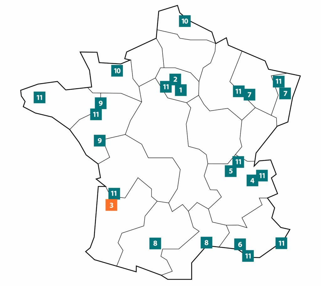
3
Bordeaux
Presentation
The Bordeaux Bio-Imaging Institute (IBIO) is a federative structure supported by the Université de Bordeaux in partnership with the CNRS and the Bordeaux University Hospital. This project has created a technological infrastructure for the study of living organisms using in vivo imaging equipment. It was set up in 2011 to support imaging research projects by academic and industrial teams from Aquitaine and elsewhere, whether or not they specialize in imaging. It is the platform of the Bordeaux node of France Life Imaging (FLI). The platform enables financial, human, administrative and technical resources to be pooled for the benefit of common objectives. The platform’s coordinating team, supported by these supervisory bodies, is made up of a coordinator, a financial manager and expert engineers dedicated to the various equipment made available. This team is at the disposal of all those who request it for any information concerning the resources made available as well as the surrounding resources.
Key imaging expertise and know-how developed as part of FLI’s scientific activities
The node’s research activity is carried out through projects linked to the various work packages of the France Life Imaging infrastructure:
- MRI contrast agents,
- Radiotracers for molecular imaging
- biological markers;
- dynamic nuclear polarization;
- Image-guided HIFU and interventional imaging;
- Mathematical simulation and modeling
Know-how :
The imaging platform offers its services at various project levels. UMS imaging experts in the various imaging modalities can contribute to the writing of imaging procedures, the construction of models including the creation of ad hoc tools (polypeptide synthesis, cell culture, animal facilities, collaboration with other platforms at the University of Bordeaux and the CHU), the production of images, their processing and post-processing and biological analyses (immunohistology; immunocytology; biochemical assays).
Laboratoires du nœud :
PTIB, UMR INCIA, UMR RMSB, Centre de recherche radio thoracique de Bordeaux, Inserm U1049, UMR 5255, LABRI
Preclinical and clinical platforms / scientific managers :
Bio-imaging platform :
- IBIO – Anne Thevenoux
- Vivoptic – Stéphane Mornet
- Life Sciences Imaging Platform – UMS 3767
4
Grenoble
Presentation
The “Imagerie des Sciences du Vivant de Grenoble” platform is dedicated to preclinical and clinical imaging research. It brings together over 120 permanent researchers, 77 PhD students and postdoctoral researchers, chemists, physicists, engineers, technologists, biologists, pharmacists and physicians. Its objective is to develop, in a harmonious and coherent way, the most relevant tools, equipment and methodologies for the in vivo exploration of physiopathological processes in animal and human models. The performance of these systems exceeds that of commercial systems. The equipment and associated expertise are available to the academic and industrial scientific community.
Key imaging expertise and know-how developed as part of FLI’s scientific activities
Methodological research focuses on (i) new optical equipment, X-ray imaging and nuclear detectors, (ii) new optical and nuclear imaging agents, (iii) intra-vital microscopy, (iv) neurophysiology, (v) MRI perfusion imaging, (vi) stereotactic radiotherapy guided by tomographic imaging. The combination of these scientific, technological and applied expertises will enable us to achieve consistency on a larger scale in integrated multimodal imaging for new in vivo molecular imaging solutions.
Preclinical and clinical platforms / scientific managers
The hub comprises 6 main imaging platforms:
- Multimodal interventional imaging ( intraoperative MRI, SPECT, MEG, confocal microendoscopy, fluorescence microscopy): Clinatec – Vincent Auboiroux, Vincent.AUBOIROUX@cea.fr
- Optical and opto-acoustic imaging : Optimal – Véronique Josserand & Jean-Luc Coll, veronique.josserand@univ-grenoble-alpes.fr, jean-luc.coll@univ-grenoble-alpes.fr.
- Nuclear imaging and US : GAIA – Alexis Broisat & Catherine Ghezzi, alexis.broisat@inserm.fr, catherine.ghezzi@univ-grenoble-alpes.fr
- Clinical and preclinical MRI, MRI-guided FUS platform, high-resolution preclinical X-ray CT scanner, X-ray CT-guided stereotactic irradiation platform: IRMaGe – Emmanuel Barbier, Tiphaine Jouanno & Hélène Elleaume, emmanuel.barbier@univ-grenoble-alpes.fr, tiphaine.jouanno@univ-grenoble-alpes.fr, helene.elleaume@inserm.fr
- MRI, Neurophysiology, EEG, TMS: iRMaGe – Aurelie Campagne, aurelie.campagne@univ-grenoble-alpes.fr
- IntraVital Microscopy – IVM : linear, non-linear and photoacoustic – Boudewijn van der Sanden, boudewijn.vandersanden@univ-grenoble-alpes.fr
Associated research laboratories
U1039, https://lrb.univ-grenoble-alpes.fr; U1209, https://iab.univ-grenoble-alpes.fr; https://www.imagerie-optimal.fr, U1216 (team 5), https://neurosciences.univ-grenoble-alpes.fr; https://irmage.univ-grenoble-alpes.fr; U1205, CLINATEC, https://www.clinatec.fr; UMR 5250, https://dcm.univ-grenoble-alpes.fr, TIMC UMR5525 (SyNaBi team), https://www.timc.fr/SyNaBi, INSERM UA7, https://strobe.univ-grenoble-alpes.fr
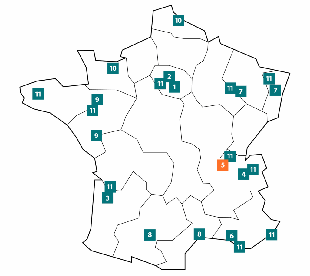
5
Lyon
Presentation
Historically, the hub was located in the Lyon region, but since the beginning of 2025, it has been expanded to accommodate the In Vivo Imaging Auvergne (IVIA) imaging platform located in the Clermont-Ferrand region.
Under the auspices of Université Claude Bernard Lyon 1 (UCBL1), the Lyon hub draws on an internationally renowned environment for biomedical and technological research in diagnostic and therapeutic medical imaging. Imaging research revolves mainly around multimodal imaging platforms with equipment installed at CERMEP on the Lyon-Est medical campus, at CREATIS on the UCBL / INSA science and technology campus, and in Clermont-Ferrand with the IVIA platform. CERMEP has been awarded the IBISA label and IVIA has been awarded the IBISA label since 2018. This node offers the scientific community a wide range of services based on its equipment park and exceptional clinical environment, complemented by a national veterinary school, VetAgroSup, hosting a research center under private contract.
The Lyon-based imaging platforms feature several innovative pieces of equipment:
- A photon-counting scanner, SPCCT, for human research, unique in France,
- One of four human PET-MRI scanners installed in France, funded by an EquipEx II and the CESAME IHU,
- High-intensity focused ultrasound (HiFU) equipment developed in labTau for MRI-guided therapy of the prostate, liver, kidneys and heart,
- One of the 5 MEGs available in France.
IVIA focuses its expertise on food and nutrition, neuroscience, cardiovascular health and oncology. IVIA is attached to the Université Clermont Auvergne (UCA), and benefits from the UCA Partner platform support structure (President: Pr Jean-Michel Chezal). It is a multi-site platform comprising 4 entities, each specialized in a particular in vivo imaging modality.
- Three units are located in Clermont-Ferrand:
– CHU Gabriel Montpied’s 3T MRI platform dedicated to humans
– the IMoST multimodal imaging platform dedicated to small animals. It brings together equipment based on different radiation sources: PET, CT, optical, multi-source SPECT-CT. The site also boasts the resources needed to carry out such studies, in particular devices for synthesizing and radiolabeling a wide range of radioactive isotopic tracers for use in imaging.
– the experimental catheterization laboratory (LCE) in the Institut Pascal’s image-guided therapy department. This entity is recognized as one of the few national interventional radiology platforms dedicated to angiography of medium-sized animals, combining ultrasound and CT. - The latest entity is located on the INRAE Theix site, around 15km from Clermont-Ferrand:
– the AgroResonance platform. It features a high-field MRI, the first 11.7 T device installed in France. This horizontal system, specially dedicated to investigations in mouse models, measures metabolic and functional signatures on different nuclei with a high level of sensitivity.
In addition, the node has well-characterized patient cohorts in neurology (Lyon Neuroscience Research Center, Stem Cell and Brain Research Institute, Cognitive Neuroscience Center, CREATIS, Stroke Research Unit), cardiology (CarMeN) and dedicated echocardiography expertise.
The research carried out in the laboratories supporting the hub’s platforms is focused on 5 main areas:
- Development and validation of dedicated imaging agents for multispectral X-ray imaging (SPCCT) for neurology, cardiovascular and oncology applications;
- Methodological developments for MRI and NMR spectroscopy
- Multimodal MRI and Intra-Vital Optical Imaging
- Digital modeling,
- Data analysis; part of the development work has made a significant contribution to the software infrastructure of the IAM node (VIP image processing environment developed within CREATIS).
Preclinical and clinical imaging platforms / scientific managers
Multi-modal and molecular imaging, PET, PET-MRI, MRI, SPCCT : CERMEP – Luc Zimmer
Center for Research in Image Acquisition and Processing for Health: CREATIS– PiLoT platform – Olivier Beuf
Ultrasound Therapeutic Applications Laboratory : LabTAU (U 1032 INSERM – UCBL) – Cyril Lafon
NMR platform for agronomy, agri-food and nutrition : AgroResonance – Jean-Marie Bonny (Auvergne Regional Manager) – Guilhem Pagés (Platform Manager) and Leslie Mazuel (Coordinator)
Research laboratories associated with the hub
Lyon:
Cardiovascular, Metabolism, Diabetology and Nutrition Research Laboratory : CarMeN – Hubert Vidal
Center de lutte contre le cancer and Hospices Civils de Lyon (HCL): Anne Metzinger
Clermont-Ferrand :
UMR 1240 INSERM/UCA, directed by Pr Elisabeth Miot-Noirault
UR370 QuaPA INRAE headed by Dr Pierre-Sylvain Mirade.
Radiology department of CHU Gabriel-Montpied, directed by Pr. P. Chabrot,
Useful links: CREATIS, Inserm U1032, IPNL, Inserm U1060, CNRL BIORAN, CNRL CMO, LAGEP , Agroresonance,
Dedicated page: Linkedin France Life Imaging – Lyon
6
Marseille
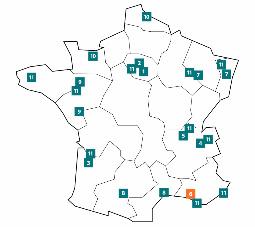
Presentation
The Marseille hub is a multidisciplinary network of high-tech imaging facilities for imaging research and applications in animal and human models. Preclinical and clinical imaging benefit from close links between specialists in instrumentation, chemistry and image processing, on the one hand, and biologists and clinical researchers on the other. This research has benefited greatly from the financial support of Investissements d’avenir (Infrastructure, Equipex, AMIDEX), ANR, foundations (James S McDonnell Foundation, ARSEP, AFM, etc.) and European contracts (M-CUBE FET-Open, IMI pharma-Cog, ERC-Gaba Networks, Neuropioneer BrainScale, Human Brain Project, Envision, Entervision, Marie Curie, ENDOTOFPET).
In total, the Marseille Node brings together 200 FTEs, including 100 permanent researchers, engineers and clinicians, dedicated to the development of in vivo imaging and its applications, in part with prestigious international partners.
The network provides access to innovative equipment for
- MRI (on the IBISA MR platform of UMR 7339 CRMBM / CEMEREM and at INT),
- Nuclear imaging and X-rays (at CERIMED, CPPM),
- Optics and biophotonics (at Institut Fresnel, INT, INMED).
Equipment for biomedical research includes 3 preclinical MRI scanners (4.7T, 7T, 11.75T), 3 multi-organ MRI systems dedicated to clinical research (1.5T, 3T, 7T), 1 3T MRI for preclinical and clinical neuroimaging, 1 PET-CT and 1 SPECT-CT for clinical research, 1 pre-clinical PET-CT and SPECT-CT, 1 pre-clinical ultrasound scanner, 1 MEG, 1 two-photon microscope, 1 bio-fluorescence imager, 1 non-linear optical endoscopy system, a non-linear optical microscope device, 1 Raman imager, 1 SPCCT pre-clinical spectral scanner.
The main research themes focus on
- The development of innovative advanced optical systems for in vivo deep brain imaging and the study of myelin organization in the spinal cord using coherent Raman imaging.
- Ultra-high-field MRI (7T) for cerebral, muscular and osteo-articular imaging, radiofrequency coils with new materials.
- The development of sodium imaging, for early detection of pathologies
- Preclinical and clinical neuro-functional MRI
- Simultaneous acquisition of MEG, EEG and SEEG signals)
- The development of a spectral X-ray scanner by the Marseille particle physics center. This system is based on a new hybrid pixel detector enabling k-edge imaging of gadolinium and gold nanoparticles. This system will enable a reduction in the dose of X-rays received by the subject, as well as molecular imaging thanks to the incorporation of gadolinium- or gold-labeled tracers into the subject. This system has been installed at CERIMED since the end of 2018 for validation.
Hub laboratories
CRMBM, CERIMED, Institut Fresnel, INMED, CPPM, INT, LIS
Preclinical and clinical platforms / scientific managers
Center de Résonance Magnétique Biologique et Médicale/exploration métabolique par résonance magnétique: CRMBM/CEMEREM UMR7339, Monique Bernard
European Research Center for Medical Imaging/Multimodal Molecular Imaging: Cerimed, Benjamin Guillet
Institut de Neurosciences de la Timone/IRMf, optics : INT UMR 7289Director Guillaume Masson, fMRI Manager, Jean-Luc Anton, Optical Imaging Manager, Ivo Vanzetta
Institute of Systems Neuroscience/Magnetoencephalography: INS UMR 1106, Director Viktor Jirsa, MEG lab – Jean-Michel Badier
Institut Fresnel/ Optics, Photonics, Electromagnetism and Signal and Image Processing, UMR 7249: Director Sophie Brasselet, Head of Optics, Photonics, Hervé Rigneault
7
Great East
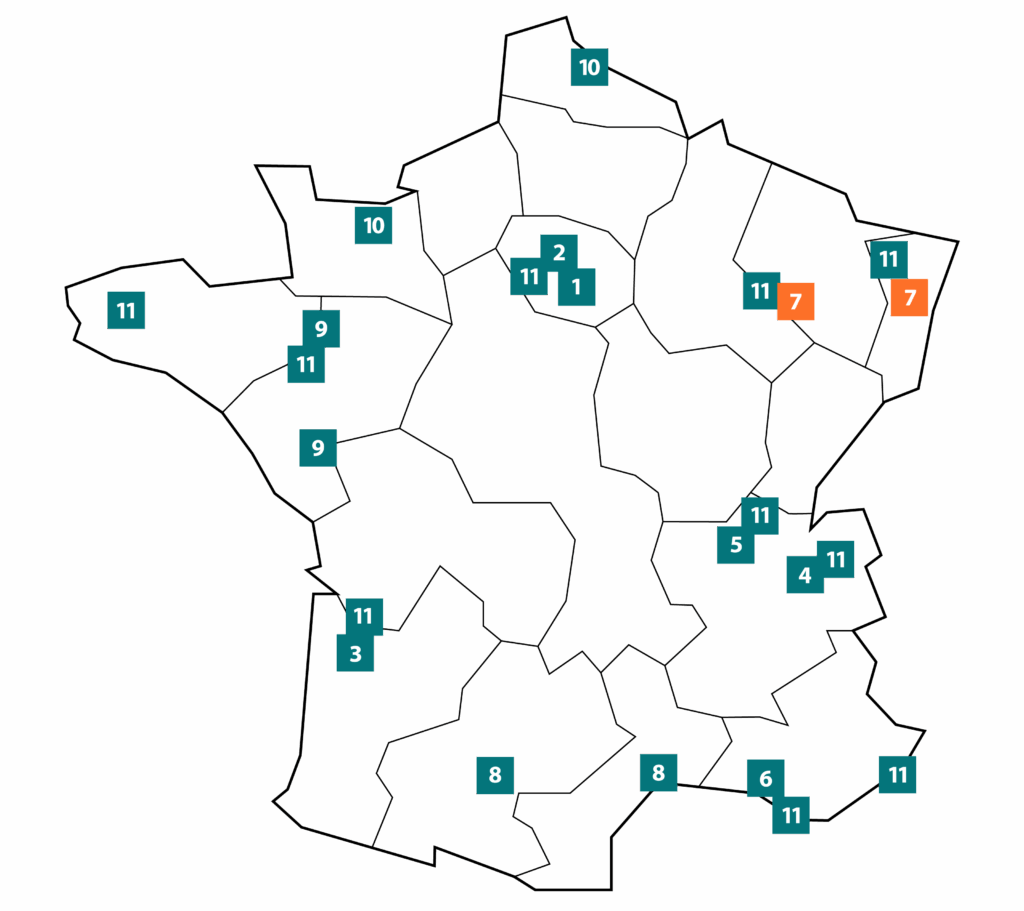
Presentation
FLI’s Grand Est hub is one of the three hubs integrated into the infrastructure on June1, 2020.
It comprises five main imaging platforms with in vivo imaging modalities available and open to the research community:
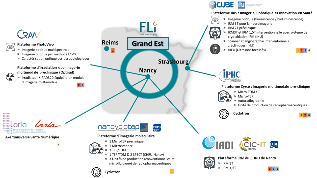
Open to academic and industrial research partnerships, the imaging facilities cover the full spectrum of in vivo imaging modalities (MRI, nuclear, ultrasound, X-ray and optical for human and animal applications). The hub boasts strong skills in instrumentation, diagnostic and interventional imaging, and robotics. These in vivo imaging platforms are complemented by strong expertise in data management, information processing and 3D navigation at the ICube laboratory in Strasbourg, LORIA-INRIA and CRAN in Nancy.
In addition, we have recognized expertise in the engineering of radiolabeling molecules for molecular imaging, combined with the production of short- and long-half-life positron-emitting radioisotopes for diagnostic and theranostic applications. This is complemented by expertise in proton beam irradiation.
Key imaging expertise and know-how developed as part of FLI’s scientific activities
The Grand Est hub has expertise in a wide range of fields:
- Functional MRI and real-time image reconstruction, movement tracking,
- Imaging the vocal tract,
- Vascular modeling and modeling of deformable organs for interventional procedures,
- 3D navigation in images to guide operations,
- Nanobiophotonics and multifunctional nanoparticles,
- Wide-field, quantitative, real-time, fluorescence and microscopic optical imaging,
- Translational research in biomedical optics (optical spectro-imaging) and multimodal panoramic imaging (oxygenation mapping),
- X-ray imaging, bioluminescence and tomography,
- HiFUs and interventional imaging,
- The development of positron-emitting molecules for molecular imaging,
- Image and data processing: 2D and 3D registration, segmentation, reconstruction, 3D navigation, feature extraction, classification,
- Brain imaging.
Preclinical and clinical imaging platforms / scientific managers :
- The IADI / CIC-IT laboratory platform in Nancy,
- The PhotoVivo platform: translational optical spectro-imaging(leaflet presenting the platform’s equipment), and the OptiRAD platform: preclinical irradiator/imager based at CRAN in Nancy,
- Nancyclotep’s molecular imaging platform in Nancy,
- The LORIA platform in Nancy,
- The IRIS platform in Strasbourg,
- The Cyrcé platform in Strasbourg.
Platform-backed research laboratories
- Laboratoire d’Imagerie Adaptative Diagnostique et Interventionnelle: IADI, INSERM U1254, Nancy,
- The Centre de Recherche en Automatique: CRAN, UMR 7039 in Nancy,
- Nancyclotep in Nancy,
- The Laboratoire Lorrain de Recherche en Informatique et ses Applications : LORIA, UMR 7503 in Nancy,
- The Engineering, Computer Science and Imaging Laboratory: ICube, UMR 7357 in Strasbourg,
- The Department of Radiobiology, Hadrontherapy and Imaging: DRHIM at the IPHC (Institut Pluridisciplinaire Hubert Curien) in Strasbourg.
8
Occitan
Presentation
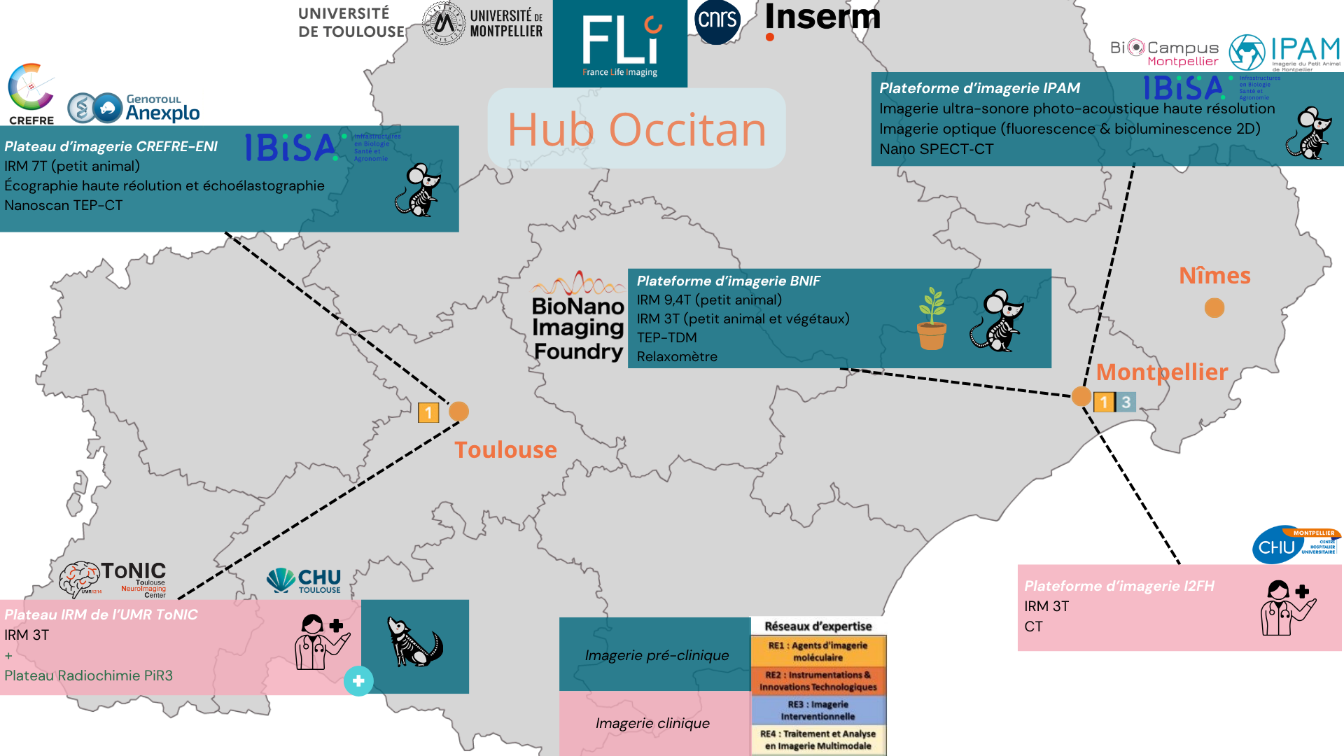
The Occitan hub joined the FLI infrastructure on June 1, 2020, at the same time as 2 other hubs.
It brings together 5 imaging platforms located in Toulouse and Montpellier, under the joint supervision of the University of Toulouse Paul Sabatier III and the University of Montpellier.
Open to academic and industrial research partnerships, our imaging facilities cover the full spectrum of in vivo imaging modalities (MRI, nuclear, ultrasound, X-ray and optical for human and animal applications).
In terms of pre-clinical imaging research, we work in Toulouse with PET-CT imaging equipment(NanoScan Mediso), 7T MRI (Pharmascan Biospec 7T Bruker MRI/MRS), ultra-fast ultrasound (Vevo2100, Visualsonics) and echoelastograph (Aixplorer Supersonic Imagine) on the CREFRE-Anexplo (Centre Régional d’Exploration Fonctionnelle et Ressources Expérimentales) non-invasive exploration platform, on the Oncopole and Rangueil hospital sites.
In Montpellier, the IPAM(Imagerie du Petit Animal Montpellier) platform of the UAR BioCampus is equipped with a high-resolution ultrasound imaging system (vevo 3100, visualsonics) and a high-resolution photo-acoustic imaging system(vevo LAZR-X, visualsonics) on the site of the PhyMedExp unit. In addition, a nano SPECT-CT imaging system (Mediso) and 2D bioluminescence and fluorescence optical imaging systems (Ivis Lumina II), on the technical platform of theIRCM (Institut de Recherche en Cancérologie de Montpellier). On the University site, the BNIF (BioNanoImaging Foundry) platform completes the imaging offering with a 9.4T MRI, two3T MRIs with vertical and horizontal magnets, and a Micro-CT. It also has a PET-CT system (Médiso), again on the IRCM technical platform.
For clinical imaging, both cities have dedicated research platforms. In Montpellier, the
Key imaging expertise and know-how developed as part of FLI’s scientific activities
The scientific expertise associated with preclinical in vivo imaging systems is mainly focused on non-invasive, longitudinal monitoring of cancer progression, to test the long-term effect of new drugs and new therapeutic targets on the IPAM/IRCM platform, but also on the study of the major physiological systems of organisms, IPAM/PhyMedExp (cardiology, neurology…). The BNIF platform boasts expertise in pre-clinical imaging on animal and plant models, with the possibility of exporting equipment and know-how directly to the field. In addition, the Toulouse site has developed expertise in oncology, metabolism, cardiovascular, infection imaging and neuroscience imaging.
In terms of clinical research, Le Node Occitan has recognized expertise in neuroscience, particularly in the field of imaging biomarkers for brain pathologies (Parkinson’s syndrome, Alzheimer’s disease, multiple sclerosis, psychiatry) and in oncology. We also have expertise in the exploration of the whole human body, both in healthy subjects and in clinical settings.
Preclinical and clinical imaging platforms / Location/ scientific leaders :
BioNanoImaging Foundry, University of Montpellier, Triolet site, Building 50
Manager: Christophe Goze-Bac
IPAM-U1046platform (ultrasound, Doppler and photoacoustics), CHU Arnaud de Villeneuve site. Manager: Pierre Sicard
IPAM-IRCMplatform (SPECT-CT and bioluminescence/fluorescence optical imaging), Institut de Recherche en Cancérologie de Montpellier site – U1194. Manager: Jean-Pierre Pouget
CREFRE-Anexplo (Regional Center for Functional Exploration and Experimental Resources)
Multimodal in vivo imaging platform dedicated to small animals, 2 sites: Oncopole & CHU Rangueil. Manager: Carine Pestourie
Montpellier University Hospital Functional Imaging Platform, Gui de Chauliac University Hospital.
Manager: Emmanuelle Le Bars
Plateau technique IRM Toulouse NeuroImagingCenter (ToNIC), Pavillon Baudot CHU Purpan.
Manager: Hélène Gros-Dagnac/Nathalie Vayssière
Research laboratories supported by the node’s platforms :
Coulomb Laboratory Montpellier University https://coulomb.umontpellier.fr/
ToNIC UMRS1214 Inserm UT3 https://tonic.inserm.fr/
UMR5549 CNRS UT3 CERCo https://cerco.cnrs.fr/les-equipes/plateau-irm/
UMRS 1037 Inserm UT3 https://www.univ-tlse3.fr/structures-de-recherche/umr-1037-centre-de-recherche-en-cancerologie-de-toulouse
IRCM U1194 https://www.ircm.fr/
Pi-R3 tray https://tonic.inserm.fr/plateaux-techniques/pi-r3-2/pi-r3/
9
Great West
Presentation
FLI’s Grand Ouest hub is one of the three hubs integrated into the infrastructure on June1, 2020.
It comprises four imaging platforms located in Rennes, Nantes, Angers and Brest: NeurInfo (Imaging and Neuroinformatics Platform), PRISM (Multi-modal Imaging and Spectroscopy Research Platform), CIMA (Applied Multimodal Imaging Center), PLaTIMed (Platform for the Design and Evaluation of Medical Devices in Brest). Members of the Biogenouest network, these platforms are supervised by the University of Rennes, Nantes University, the University of Angers, INRAE and the University of Bretagne Occidentale. Imaging facilities open to academic and industrial research cover the full spectrum of
For preclinical research, the hub has the following equipment:
– a 7T MRI located in Angers, a 4.7T MRI located in Rennes, dedicated to small animal imaging and spectroscopy, and a 1.5T MRI located in Rennes, for medium animal imaging (pig model), as well as two high-resolution NMR devices on the PRISM platform(https://www.pf-prism.org/les-equipements/).
– nuclear imaging equipment dedicated either to the average animal (miniature pig) – 1 PET-CT, 1 CT, and 2 gamma cameras on the PRISM platform(https://www.pf-prism.org/les-equipements/)– or to laboratory rodents, with a PET-MRI, a PET-CT and a unique 3-photon PET device at CIMA in Nantes(https://crci2na.univ-nantes.fr/en/research/cima). Both platforms also have radiopharmaceutical equipment for the synthesis of radiolabeled molecules.
– computed tomography devices(https://platimed.fr/)
Clinical research relies mainly on MRI and computational models implemented on the NeurInfo platform(https://www.neurinfo.org/). Neurinfo is equipped with a 3T MRI installed at Rennes University Hospital.
Key imaging expertise and know-how developed as part of FLI’s scientific activities
Thanks to these facilities, the Grand Ouest node has expertise in the following areas:
- Quantification of physiological and metabolic parameters (perfusion, concentration of molecules of interest, body composition, receptor density) and other biomarkers (water diffusion, relaxation time, etc.) for preclinical applications in oncology and nutrition (PRISM platform),
- Preclinical molecular imaging applied to immunology and oncology,
- image processing and analysis in the context of oncology
- tomographic image reconstruction
- Neuroinformatics in the context of nervous system diseases,
- Population imaging.
Preclinical and clinical imaging platforms / scientific managers :
- PRISM(https://www.pf-prism.org/les-equipements/)/ Pierre-Antoine Eliat
- CIMA in Nantes (https://crci2na.univ-nantes.fr/en/research/cima)/ Michel Cherel
- NeurInfo (https://www.neurinfo.org/fr/)/ Emmanuel Caruyer
- PLaTIMed(https://platimed.fr/) / Guillaume Dardenne
Research laboratories supported by the node’s platforms :
- UAR 3480 CNRS, US18 INSERM, Biosit (https://biosit.univ-rennes.fr/)
- INRAE 1341, INSERM 1317, Institut NuMeCan, Equipe EAT(https://numecan.fr/eat/)
- UMR INSERM 1099, LTSI(https://ltsi.univ-rennes.fr/)
- UMR INSERM 1066 – CNRS 6021, MINT(https://mint.univ-angers.fr/en/index.html)
- INSERM UMR1307 – CNRS UMR6075 – CRCI2NA (Team 2) (https://crci2na.univ-nantes.fr/en/research/team-2)
- UMR CNRS 6074, ERL U1228 Empenn(https://team.inria.fr/empenn)
- UMR INSERM 1101 UBO IMT Atlantique LaTIM(https://latim.univ-brest.fr/ )
10
North West
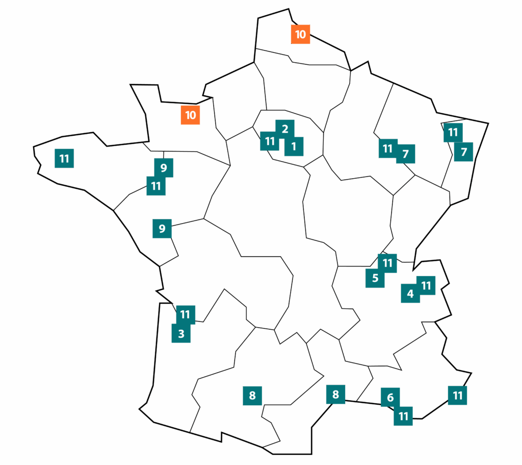
Presentation
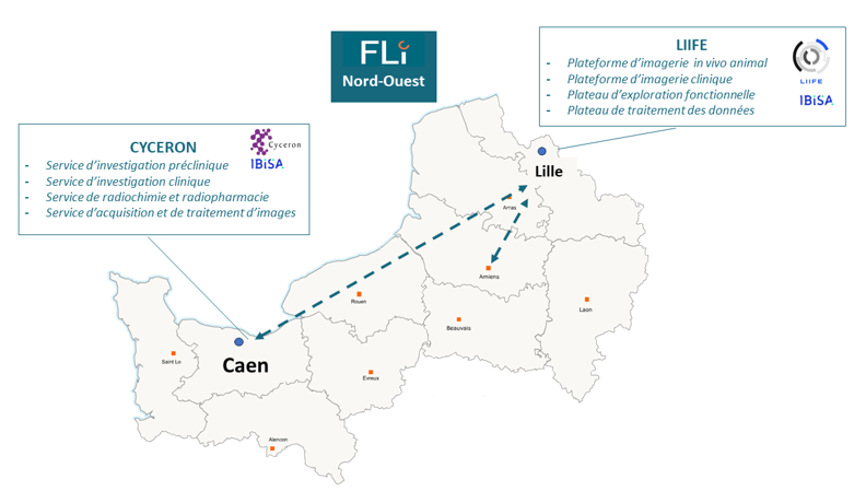
The North-West Hub is the new hub of France Life Imaging (FLI), integrated into the infrastructure on January 1, 2025. It brings together two major IBISA-certified multimodal imaging platforms:
- LiiFE (Lille In vivo Imaging and Functional Exploration) in Lille(https://ums-plbs.univ-lille.fr/les-plateformes-constitutives/imagerie-du-vivant-exploration-fonctionnelle)
- Cyceron in Caen(https://www.cyceron.fr/index.php/fr/).
These platforms are supervised by :
- INSERM, Université de Lille, Institut Pasteur de Lille and CHU de Lille for LiiFE,
- Inserm, CNRS and the University of Caen for Cyceron.
The Lille and Caen sites offer integrated in vivo imaging modalities and experimental models for a better understanding of the physiology and pathophysiology of human diseases, including neurology, psychiatry, oncology and cardiology.
State-of-the-art for preclinical and clinical research
The Hub Nord-Ouest provides a multimodal imaging park with state-of-the-art equipment:
In Lille :
Clinical research :
- 7T MRI dedicated to research (shared with Amiens University Hospital)
- 3T MRI
- OCT Spectralis Heidelberg
Preclinical research :
- µ-IRM 7T dedicated to rodent imaging
- µ-PET-CT dedicated to rodent imaging
- 0.2T MRI for large animal imaging
- A functional and behavioral exploration platform
In Caen :
Clinical research :
- 3T MRI + EEG MRI compatible
- PET-CT
Preclinical research :
- µ-IRM 7T
- µ-MRI 7T-PET with cryoprobe
- 2 X 7T MRI
- PET-CT
- µ-PET-CT dedicated to rodent imaging
- 3T MRI and PET-CT for large animals
- MPI (Magnetic Particle Imaging) for mice (unique in France)
- fUS for small and large animals
- Angiograph X
- X-ray irradiator
- Cleanroom for the production of radiopharmaceutical agents
Ae expertise in biomedical imaging
The Hub Nord-Ouest offers advanced integration of in vivo imaging modalities and experimental models to improve understanding of human physiology and pathology.
Areas of expertise :
- Development of experimental models
- Innovative multimodal imaging.
- Radioisotope production and chemistry for imaging.
- Expertise in cerebral, cardiac, oncological and retinal imaging.
- Functional and behavioral investigations.
- Image acquisition, harmonization and processing.
- Artificial intelligence applied to imaging.
- Scientific promotion and project support.
Preclinical and clinical imaging platforms / Location / Scientific leaders :
The platforms are open to collaborations and imaging research services with academic and industrial partners.
- Plateforme LIIFE – Lille In vivo Imaging and Functional Exploration (20 people)
Campus du CHU de Lille, UFR 3S, Pôle Recherche
Heads: Pr. Jean-Pierre Pruvo and Dr. Renaud Lopes
https://ums-plbs.univ-lille.fr/les-plateformes-constitutives/imagerie-du-vivant-exploration-fonctionnelle - CYCERON platform (50 people)
Campus EPOPEA, Caen
Responsible: Dr. Benoit Haelewyn
https://www.cyceron.fr/index.php/fr/
Research laboratories supported by the node’s platforms :
Lille :
- U1172 LillNCog “Lille Neuroscience & Cognition”(https://lilncog.eu/)
- UMR 1011 “Nuclear Receptors, Metabolic and Cardiovascular Diseases”(https://pasteur-lille.fr/centre-de-recherche/unites-de-recherche/recepteurs-nucleaires-maladies-metaboliques-et-cardiovasculaires/)
- U1008 “Advanced Drug Delivery Systems”(https://u1008.univ-lille.fr/)
- U1189 OncoThai “Laser-assisted therapies and immunotherapies for oncology”(https://www.oncothai.fr/fr/component/tags/tag/onco-thai)
- UMR 1003 PhyCell “Laboratory of Cell Physiology”(https://phycell.univ-lille.fr/)
- UMR 9020 CNRS UMR1277Inserm Canther “Cancer Heterogeneity Plasticity and Resistance to Therapies”(https://canther.fr/en/canther-cancer-heterogeneity-plasticity-and-resistance-to-therapies/)
- UMR 1190 RTD “Translational Research in Diabetes”(https://www.translational-research-diabetes.com/FR/index)
- UMR 1283 / 8199 “Functional (Epi)genomics and Molecular Physiology of Diabetes and Related Diseases”(https://www.good.cnrs.fr/)
- U1286 INFINITE “Institute for Translational Research in Inflammation”(https://lille-inflammation-research.org/fr/)
- Inserm U1019 -CNRS UMR9017 CIIL “Centre d’infection et d’immunologie de Lille”(https://www.ciil.fr/fr/)
- ULR 4483 – IMPECS “Impact of the chemical environment on health”(https://impecs.univ-lille.fr/)
Caen :
- UMR 1237 PhIND “Physiopathology and Imaging of Neurological Disorders”(https://www.phind.fr/index.php/en/)
- UMR 1077 NIMH – BB@C “Neuropsychology and Imaging of Human Memory”(https://nimh.unicaen.fr/fr/accueil/)
- UMR 1075 COMETE “Mobilités: Vieillissement, Pathologies, santé”(https://comete.unicaen.fr/)
- UMR 6030 ISTCT “Imagerie & Stratégies Thérapeutiques pour les Cancers & Tissus Cérébraux”(https://www.istct.cyceron.fr/index.php/fr/)
- UMR 8240 LaPsyDÉ “Laboratoire de Psychologie du Développement et de l’Éducation de l’Enfant”(https://www.lapsyde.com/)
- UR 4650 PSIR “Physiopathology and Imaging Strategy of Cardiovascular Remodeling”(https://www.unicaen.fr/laboratoire/ur-4650-physiopathologie-et-strategies-dimagerie-du-remodelage-cardiovasculaire-psir/)
11
IAM “Information Analysis & Management
Presentation
The “Information Analysis and Management” hub, IAM(https://portal.fli-iam.irisa.fr/home), is tasked with building an infrastructure (hardware and software) accessible to users of national in vivo imaging platforms, in order to facilitate the opening up and reuse of imaging data, and to store, manage and process large sets of clinical and preclinical imaging data and associated metadata.
The hub brings together medical image processing experts from French node and non-node laboratories, under the umbrella of INRIA (Rennes), CNRS (ICUBE, I3S, Creatis, CRMBM), CEA (MIRCen) and INSERM (Brest, Nancy, Grenoble). Scientific coordination of the node has been entrusted to INRIA. Initially coordinated by a tandem formed by Christian BARILLOT and Michel DOJAT assisted by Michael KAIN, the IAM hub has been coordinated by Michel DOJAT with the support of Michael KAIN since Christian BARILLOT passed away in June 2020. We are grateful to our colleague Christian for the work he has accomplished over the years, for his ability to bring people together and for his vision.
The project was implemented in two stages. The first, from 2013 to 2018, was devoted to the creation of a federated image analysis and data management solution that would ensure the interoperability of existing heterogeneous and distributed solutions implementing raw and metadata indexing (e.g., using semantic models or ontologies) of information. Four pre-existing national IAM management solutions were selected: CATI (CEA, Paris-Sud), SHANOIR (INRIA, Rennes), MediBase (CNRS, Strasbourg) and Archimed (INSERM, Nancy). Three existing national solutions for image processing and workflow management were initially selected: VIP (CNRS, Lyon), MedInria (INRIA, Rennes) and BrainVisa (CEA, Paris-Saclay).
The IAM node set up a steering committee to define the node’s action priorities and orientations. A technical manager (Mr. Kain) supervised the developments carried out by a team of up to 15 software engineers based in six different centers. In order to provide the most comprehensive response possible, three working groups were set up:
- The first dealt with interoperability issues, in particular interoperability between data repository solutions.
- The second dealt with image processing problems in order to provide versatile solutions for parallel or massive computing.
- The latter focused on transferring the existing clinical imaging environment and expertise to the emerging preclinical imaging sector.
Since November 2020, the industrialization and operation of the software platform has been entrusted to a temporary consortium of companies, selected following a call for tenders. The contractor offers services based on the data management and processing platform developed by IAM, and the deployment of processing chains on high-performance computing systems to handle large databases.